The Aberdeen Centre for Electron Microscopy, Analysis and Characterisation (ACEMAC) is a facility open to electron microscopy users from both across the university and beyond. Dr Alex Brasier is the academic lead at the Centre and a Senior Lecturer in Geology at the University of Aberdeen, working on carbonate rocks (limestones). He uses electron microscopy (including the Centaurus CL detector from Deben) for both academic research and teaching.
Dr Brasier talks about his use of cathodoluminescence detectors and why he chose to purchase the Centaurus from Deben. “Cathodoluminescence is a technique commonly used on carbonate rocks to look at crystal growth histories. Using the other detectors of the electron microscope, I can see crystal sizes and shapes as they appear today. What the CL detector gives is an image of what the limestone looked like prior to alteration of crystals such as by continued crystal growth or nanometer-scale dissolution and re-precipitation of the crystal. This is possible because CL patterns reflect the distribution of trace elements like manganese and iron in the crystal, and these trace element patterns can be unaffected by such rock-altering ‘diagenesis’. In effect the CL reveals ‘ghosts’ of the original crystals that made up the rock. I have also used the CL detector for looking at growth histories of quartz crystals, and the technique is similarly useful for looking at crystals of zircon (zirconium silicate) that are commonly targeted for U-Pb age-dating.”
Continuing, Dr Brasier says “I still regularly use a traditional cold cathode optical CL system as this is also very useful for looking at carbonate rocks, but it reveals different things from the SEM CL detector. With optical CL, you see colours that relate to concentrations of particular trace elements and this can help identify different mineralogies or phases of crystal growth. With the Deben CL detector, the images are greyscale but can easily be related to backscatter electron images and energy dispersive spectrometry (EDS) X-ray element maps of exactly the same part of the same specimen. Having the CL on the SEM also means that smaller sample areas can be targeted than is possible with an optical CL (commonly I’m looking at zonation within individual crystals with the SEM CL). We tried a few proposed in-house solutions from electron microscope manufacturers, but none of these produced a satisfactory image with carbonate rocks. We knew other SEM labs had a Deben Centaurus CL detector so we sent some specimens away for a test, and the results were much better. The issue with unsuitable detectors is usually ‘streaking’ of bright stripes across the image.”
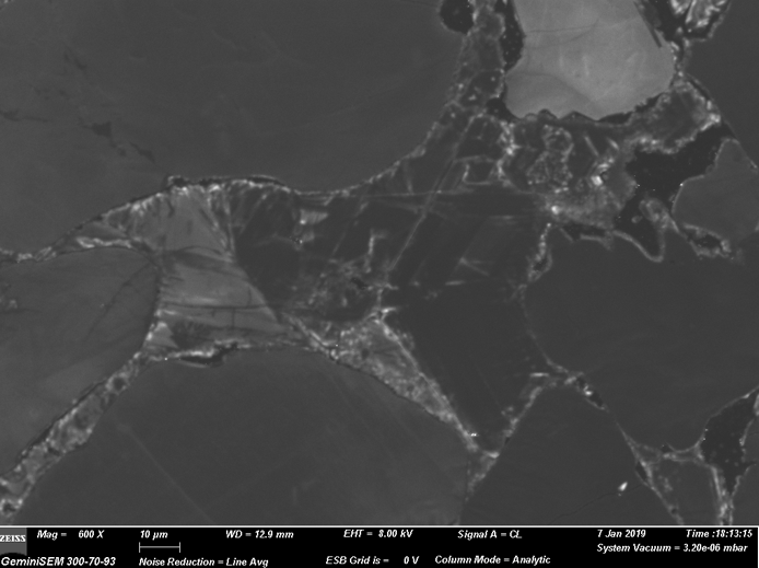
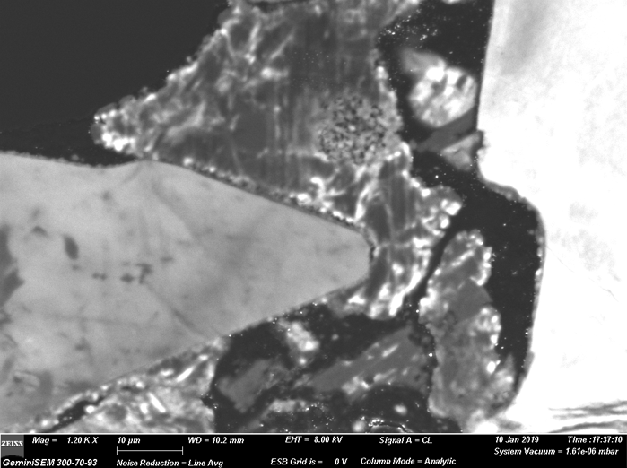
Cathodoluminescence images showing framboidal microcrystalline iron-oxide aggregates in thin section emphasising the power of the Deben Centaurus CL detector – no streaking!
You should hear from us shortly.

