![]()
In many applications, understanding response to mechanical stress is critical to better understand material behavior. While performing mechanical characterization on a sample, it can be very challenging to observe its three dimensional structure evolution without interrupting the test and preserving the part. Non Destructive Testing (NDT), such as acoustic, electromagnetic, laser or thermal, are solutions to characterize a material without altering the tested sample. However, it is very challenging to perform data acquisition while constraining the specimen because NDT techniques usually involve to release the part from external components during data acquisition.
In-situ x-rays computed tomography gives access to three dimensional information during mechanical test thanks to the integration of a mechanical stage directly inside a CT system. This technique has become a state of the art in lab microCT experiment, but how does it work exactly and how complicated is it to run such experiments ?
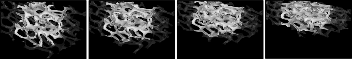
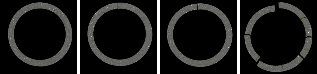
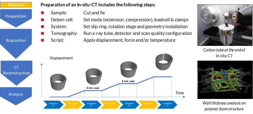
As an example of the user-friendliness association between Deben and RX Solutions systems, two samples have been studied through in-situ-CT. The first specimen is a carbon fiber tube which has been broken under a monitored displacement. Cracks creation and propagation can be observed and give useful information on its mechanical resistance. The second one is a polymer foam structure which has been compressed to observe its geometrical deformation.
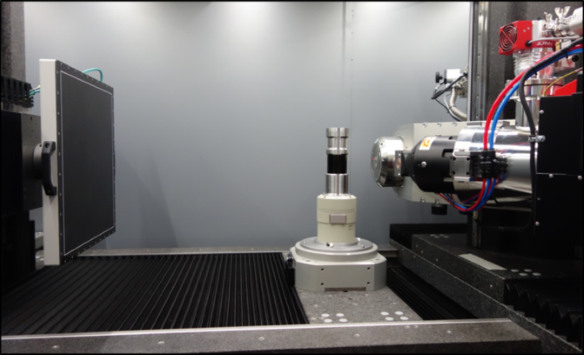
To perform these two in-situ-CT, a CT5000 RT Deben cell has been used in its compression mode with a 5kN loadcell and compression plattens. This device has been integrated into the RX Solutions EasyTom XL Ultra CT scanner equipped with a granite table, a large flat panel, an accurate air bearing rotation stage and two Xray tubes. This setup covers a very large range of resolution down to 300nm and offers a very large volume to setup samples and equipment needed.
Additionally, a specific mounting adaptor with a slip ring facilitate the Deben cell integration inside the system. It is then simply placed on the rotation stage as a standard sample holder and does not need any additional connection. Unlimited numbers of turns are then possible, which enables continuous rotation during the test.
CT Reconstruction is completed with RX Solutions proprietary XAct software which contains geometry corrections, artefact reduction algorithms (ring, beam hardening, metal, phase contrast…), filtered back projection method and batch reconstructions of multiple acquisitions. Multiple parameters can then be analysed such as, wall thickness, cell volume, void/inclusion ratio and crack propagation. Once parameters are chosen, automatic analysis can be performed on each reconstructed volume.
By automating the process, linking XAct and Deben proprietary MICROTEST software the complete acquisition sequence is automatic. Using Digital Image Correlation (DIC), it is also possible to get displacement, stress or strain fields all along the experiment from 2D Xray images or 3D CT reconstructions. These results are obtained without releasing the sample from the beginning to the end of the test.
Fatigue cycles can also be implemented and three dimensional volumes easily reconstructed at different steps in order to observe step deformations.
Thanks to the wide range of results that can be provided, in-situ X-ray computed tomography has many applications in various fields, among them can be listed the following:
In-situ-CT tests presented here requires loading to remain constant during CT acquisition to obtain a non blurred volume. However, with faster and faster scans possible thanks to new CT system development, 4D-CT might be the next step for in-situ-CT, getting closer to work which now has to be carried out at a Synchrotron.
You should hear from us shortly.

