Dr Jing Du is an assistant professor in the Department of Mechanical & Nuclear Engineering at the Pennsylvania State University (PSU). She specialises in the use of X-ray tomography (CT) to characterise advanced bio and composite materials. Her research focuses on the bio-inspired design, advanced fabrication and material characterization of medical or energy related materials and structures, such as biomedical devices inside the human body, biological organs and thin-film organic electronic devices. The work combines the experimental characterization of structures with analytical and computational models that enable the prediction of mechanical behaviour at the micro and macro-scales.
Dr Du describes her work applying CT. “We have received an award from Oak Ridge Associated Universities (ORAU) to use the Deben system to explore the mechanics of polymer-ceramic composites. (https://www.mne.psu.edu/News/2017/17-JingDuORAU.aspx). My research will examine three-dimensional deformation, strain and microstructure of polymer-ceramic composites, which can be used in tissue engineering scaffolds, capacitors, piezoelectric transducers, and many other applications. In our prior work, we used polymer-ceramic composites for bio-inspired design of functionally graded dental restorative materials. There is a need to study their mechanical properties, microstructure and the structure-properties relations for the design of robust materials and the design of robust structures using these materials. We have also received a pilot grant from National Center for Advancing Translational Sciences (NCATS) at the National Institutes of Health (NIH) through Penn State Clinical and Translational Science Institute (CTSI) to continue our implant mechanics work, again using the Deben CT5000 stage.”
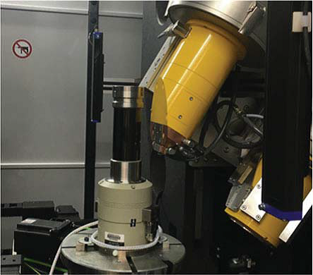
Deben CT5000 stage mounted in an x-ray tomography system from GE.
Dr Du describes how she chose to buy a Deben stage over other options. “After considering another couple of vendors, I decided to continue with Deben. I had used it during my post-doctoral studies with Professor Sunita Ho at the University of California San Francisco. I like working with Deben for several reasons. Their CT5000 is versatile and can be used in many CT systems. The technical support from Deben is very strong and very responsive. The support technicians are willing to listen to our non-traditional needs for scientific research. They are willing to modify their equipment and even make new designs. They provide strong support for our exploration of new science and technology.”
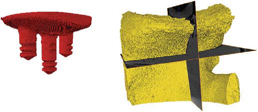
Rendered 3D images of a shoulder implant (left) and a shoulder bone (right) obtained from segmentation of micro-CT images
(images produced with permission of the authors; © The Minerals, Metals & Materials Society 2018)
An example of Dr Du’s group’s work was presented at TMS 2018, the 147th annual meeting of the Minerals, Metals and Materials Society1. Here, X-ray tomography techniques were applied to explore the damage to bone and the loosening of implants, specifically to study the deformation of shoulder bone under various physiologically realistic loading conditions.
To obtain full details of the Deben applications and product portfolio, please visit www.deben.co.uk
Reference
1 3D Full-Field Mechanical Measurement of a Shoulder Bone Under Implant Loading, Yuxiao Zhou et al, The Minerals, Metals & Materials Society, TMS 2018 147th Annual Meeting & Exhibition Supplemental Proceedings, The Minerals, Metals & Materials Series,
https://doi.org/10.1007/978-3-319-72526-0_26
You should hear from us shortly.

